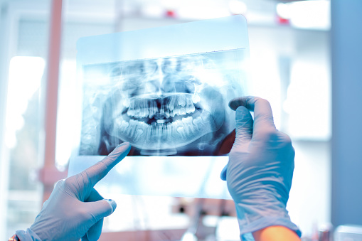
DENTAL X-RAY
AUSTIN, TX
Prior to surgical treatment, we will want to evaluate and gain a full understanding of the tissue and surrounding oral structure. This often includes taking a closer look with the use of digital x-rays.
Many patients are familiar with the standard 2D dental x-ray, in addition we can create both 3D and panoramic digital x-ray images to plan treatment and create a precise map with the entire structure being taken into consideration.
Working with our staff at Oral Surgery Specialists of Austin, we provide a thorough workup when discussing treatment, including gaining a deeper understanding with digital technology.

While much of your oral structure is visible, there is a lot that is not. Digital radiographs images allow us to view the parts that we cannot see including the roots of your teeth, impacted teeth, and your jawbone. Oftentimes, these hidden structures are essential when diagnosing issues and discussing treatment options. Digital x-rays can be categorized into four areas including:
Periapical Digital X-Ray
Periapical are the standard 2-D dental x-rays that you are probably most familiar with. They are a great tool for basic, general viewing of the inside layer of a specific tooth. The x-ray tool is mounted near your chair in the treatment area. Today, these images are taken digitally, they no longer require chemicals or processing. The images are taken quickly and viewed immediately on the computer screen nearby.
Digital technology has made the basic dental x-ray easy to capture, easier to see, while using significantly less radiation.
Panoramic Digital X-Ray
A panoramic digital x-ray image is also a 2-D image, but instead of a small area, it is designed to capture the entire mouth in a single image. It creates a flat , 5″ x 11″ image that includes a view of the upper and lower jawbone, the teeth and their roots, along with the surrounding structures and tissues.
We may use a panoramic x-ray to reveal dental and medical problems including:
- Impacted Teeth
- Jaw disorders, including TMJ dysfunction
- Advanced Periodontal Disease
- Cysts
- Sinusitis
- Tumor development or oral cancer
Cephalometric Digital X-Ray
A cephalometric digital x-ray captures a side view x-ray of the entire head. This image is used to look for alignment issues in the jaw and bite related issues.
3-D Images
3-D imaging captures considerably more than a 2-D x-ray, and is frequently used in surgical planning. Cone Beam technology provides digital tomographic 3D images. This means that we can have a complete view including depth. Having a full 360-degree view of a tooth and the entire oral cavity allows for precise mapping. all surrounding areas.
Three dimensional images can be used for customized services including bone grafts, the surgical planning for dental implants, and root canals. Having a digital reconstructed image of your skull allows for a full understanding of the oral cavity.
SURGICAL PLANNING WITH DIGITAL TECHNOLOGY
We use the advancements in technology to provide better care for our patients, including in radiography. For more information, contact our Austin office at (512) 351-7653.
More Dental Services
3D Imaging
Facial Trauma Surgery
Oral Cancer Surgery
Single Tooth Dental Implants
Anesthesia
Full Arch Replacement with Dental Implants
Oral Pathology
Sleep Apnea Treatment
Bone Graft
Impacted Canine
Implant Anchored Dentures
Pre-Prosthetic Surgery
TMJ Treatment
Cleft Lip and Palate
Impacted Wisdom Teeth
Jaw Surgery
Ridge Augmentation
Tooth Extraction

